Next: P-selectin
Up: Molecules involved in leucocyte
Previous: Molecules involved in leucocyte
Index
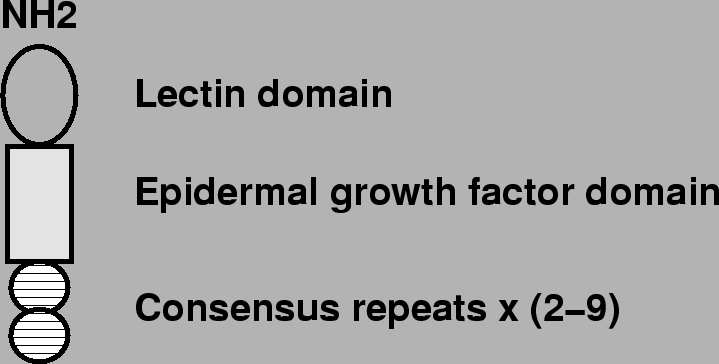
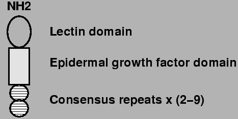
Figure 3:
Schematic representation of the molecular structure
of Selectins. On the amino terminal there is a Lectin like domain. There is
also an epidermal growth factor like domain and 2-9 consesus repeats as are
found in complement regulatory proteins.
 |
There are three members of the selectin family that participate in
``rolling''. These are P,E and L-selectin. They are expressed on the surface
of leucocytes and activated endothelial cells. Selectins have a lectin domain
on the amino (NH ) terminal, an epidermal growth factor like domain and a
series of two to nine short consensus repeats similar to complement regulatory
proteins, see figure 3.
) terminal, an epidermal growth factor like domain and a
series of two to nine short consensus repeats similar to complement regulatory
proteins, see figure 3.
The selectin that plays a central role is P-selectin.
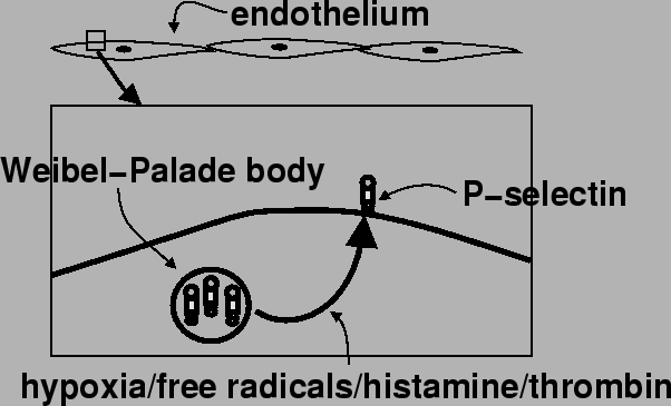
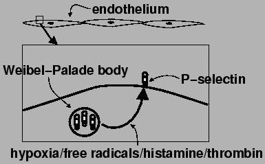
Figure 4:
Schematic representation of life cycle of P-selectin. It
is manufactured and stored in the Weibel-Palade body in endothelial cells (it
is also found in  granules in platelets). Stimulation by hypoxia, free
radicals, histamine and thrombin results in expression of P-selectin on the
surface of the endothelial cell. The main ligand of P-selectin appears to be
sialated Lewis on Leucocytes. It may also bind with L-selectin on leucocytes.
granules in platelets). Stimulation by hypoxia, free
radicals, histamine and thrombin results in expression of P-selectin on the
surface of the endothelial cell. The main ligand of P-selectin appears to be
sialated Lewis on Leucocytes. It may also bind with L-selectin on leucocytes.
 |
Subsections
Next: P-selectin
Up: Molecules involved in leucocyte
Previous: Molecules involved in leucocyte
Index
Adrian P. Ireland