Colo-Rectal

Last updated (26 October 2003)

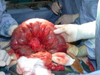
Distended Colon Click on the image to download a larger version
The clinicians were convinced about the diagnosis of toxic colitis, neither CT scanning nor colonsocpy were performed and the patient brought to theater.
What does this intra-operative image show?
Normal colon
Tumour of the transverse colon
Transverse colon grossly distended with gas
Volvulus of the transverse colon
The greater omentum has been separated from the trasnverse colon

The answer

The image shows a grossly distended transverse colon, from which the greater omentum has been separated.

Small Print

This colon is not normal. It appears grossly distended.

There is no evidence of a tumour on this image. One may see a collapsed segment distal to an obstructing tumor or a deformed area where a tumour lies.

The transvere colon is grossly distended with gas. Bowel becomes distend with gas, fluid, food debris or a combination.

There is no evidence of a volvulus of the transverse colon on this image. Volvulus indicates that the bowel is twisted around itself. It is commonest in the sigmoid colon. You may also rarely see a caecal volvulus, but volvulus of the transverse colon is extreemly uncommon.

The greater omentum is seen to lie over the patients chest in the image. Normally the omentum is attached to the transverse colon.


Adrian P. Ireland