| Good Stuff |
| Spot Diagnosis |
| Biliary System |
|
|
|
|
|
|
|
|
|
|
|
|

|
Click on the image to download a larger version |
|
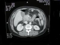
Do you see anything abnormal?
|
|
Biliary System |
| Last updated (28 October 2003) |
|
|
Signs of severe pancreatits would be lack of consistant enhancement and areas that do not enhance suggest ischemia and necrosis. Other worrysome features would be gas flecks in the pancreas. Ascitis is common in pancreatits.
A word of warning,
The head of the pancreas appears enlarged and there is fat stranding best seen in the retroperitoneum in relation to the head of the pancreas and extending into the fat between the inferior vena cava and liver and here (pointing) between the right kidney and liver.
There is a lucency in the head of the pancreas just medial to the duodenum which may represent a dilated common bile duct.
I dont see evidence of ascitis, or lack of enhancement in the head of the pancreas, there is no focal fluid collection or free gas seen.
The patient most likely has pancreatits, the enlarged bile duct raises the possibility of this being related to gallstones.