Image dscn1066 |
| Last updated (28 October 2003). |

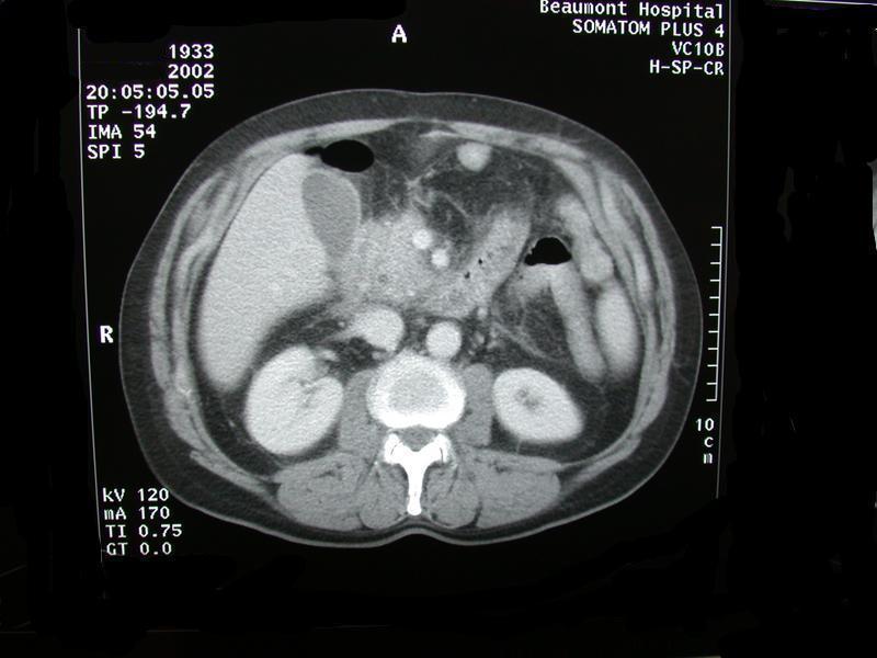
This image shows a section from an abdominal CT scan.
See notes on the usual orientation of CT scans.
This image is a section from an abdominal CT scan around the level of the epigatrium.
The date of birth is noted as 1933. The date of the scan is 2002. The patient was therefore about 69 when the scan was taken.
Intravenous and oral contrast have been given. The presence of intravenous contrast can be seen from the increased density seen over the blood vessels. The presence of oral contrast can be seen as the increased density seen in the loops of bowel.
The head of the pancreas is enlarged, there is stranding in the fat extending from the head of the pancreas into the retroperitoneum between the liver and the right kidney. There is a lucent area in the head of the pancreas raising the possibility of a dilated common bile duct.