Imagemap - dscn1066

Last updated (28 October 2003).

Aorta enhanced with iv contrast Inferior Vena cava enhanced with IV contrast Superior Mesenteric vein enhanced with IV contrast Superior Mesenteric Artery enhanced with IV contrast Date the scan was taken Patients date of birth Right kidney enhanced with contrast Branch of right renal artery The right renal vein Left kidney enhanced with contrast Gerotas fascia surrounding the right kidney Gerotas fascia surrounding the left kidney Bile duct in the head of the pancreas Vertebral body Right vertebral lamina Left vertebral lamina Posterior spine of vertebra Articular process of vertebra Articular process of vertebra The spinal cord Duodenum enhanced with oral and IV contrast Duodeno Jejeunal flexure Head of the Pancreas Fat stranding between liver and right renal vein The liver enhanced with IV contrast The lumen of the gallbladder The wall of the gallbladder The right psoas muscle Left psoas muscle Right erector spinae muscle Left erector spinae muscle Gas in the colon Small intestine enhaced with oral and IV contrast The retroperitoneum Right rectus abdominus muscle Left rectus abdominus muscle Right external oblique muscle Left external oblique muscle Right internal oblique muscle Left internal obliqe muscle Right transversus abdominus muscle Left trasnversus abdominus muscle Marker indicating the patients right hand side

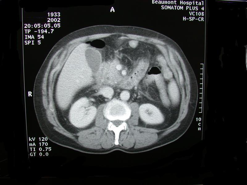
Move your pointer around the image to name the parts. Where there is some information your pointer should change from an arrow to a hand. A pop up box with information should appear if you take your hand off for a moment. If that does not work try clicking.


Adrian P. Ireland