The answer
Orientation of the patient and scan
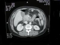
Note the vertebral column lying posteriorly. The scan should be viewed as an
axial section through the patients epigastrium with the patient lying supine,
the head is in the background and the feet the foreground. The right side of
the patient body's is seen to the left of the image and the left side of the
patients body to the right. The upper part of the image corresponds to the
patients front (anterior or ventral) and the lower part of the image
corresponds to the patients back (posterior or dorsal). This is the usual
orientation for CT body scans.
While you are studying the image you could say;
Here is the patients back (pointing), here the front, here the
right side and here the left. This is the vertebral column.
Display your advanced anatomical knowledge
The oval dense structures on each side of the vertebral column are the kidneys.
They appear with the same density as the vertebral column (iso-dense) due to
the intra-venous contrast agent that has been administered to the patient.
Surrounding each kidney is the fat of Gerota's fascia. It appears much less
dense than the kidney and other organs. However, note the pitch black areas in
the anterior abdomen. This corresponds to gas.
Here is the right kidney (pointing), and here the left.
The large structure in the right upper quadrant anterior to the right kidney is
the liver. It appears denser than it would in a scan without contrast. The
liver does not appear as dense as the kidneys because more contrast agent is in
the kidneys than the liver. There is more contrast agent in the kidneys because
of the higher arterial blood flow to the kidneys (25% of the cardiac output,
while 75% of the blood supply to the liver comes in the portal vein).
The globular structure under the right lobe of the liver, which is
less dense than the liver is the gallbladder. In this case the spleen is not
visible, either the scan is too low to see the spleen or the spleen is not
there. Just medial to the lower part of the gallbladder is a loop of intestine, probably the duodenum.
This is the liver (pointing). And this is the gallbladder
There are four nice circular densities in the middle region of the abdomen
medial and anterior to the kidneys, they apper to be paired with the largest
pair posterior and the smaller pair anterior.
The posterior pair represent the aorta and inferior vena cava. The anterior
smaller pair represent the superior mesenteric artery and vein.
The aorta is normally on the left side of the abdomen and the inferior vena
cava on the right.
The superior mesenteric vein is lateral to the superior mesenteric artery. The
portal vein is the continuation of the superior mesenteric vein when the
splenic vein which travels behind the pancreas joins the superior mesenteric
vein. Here we see a large distance between the aorta and the mesenteric vessels
which are traveling anterior to the pancreas, therefore this could not be the
portal vein, it must be the superior mesenteric vein.
This is the aorta (pointing), here is the inferior vena cava.
Here are the superior mesenteric vessels anterior to the
pancreas, this is the superior mesenteric artery and this the
vein. This structure between the aorta and the superior
mesenteric vessels is the pancreas
