The best position for the patient is in the prone Jack-Knife position. This is harder to organise than the lithotomy position and some surgeons perform the operation in the lithotomy position to avoid the difficulties in placing the patient in the prone Jack-Knife position.
Prepping and draping of the perineum come next, some surgons advocate shaving off hairs, others do not.
The surgeon places two digits of his gloved hand into the anal canal and then inserts a bivalved anal speculum or a slotted proctoscope. The surgon carefully plans out the procedure at this stage, one of the main aims is to maintain wide muco-cutaneous bridges to maintain continence and to prevent post-operative anal stenosis.
A long acting local anaesthetic combined with dilute adrenaline 1:100,000 is injected into the haemorrhoids to be excised. The surgon then waits for a few minutes for the adrenaline to have some effect and for some of the injected fluid to be absorbed.
The surgeon removes the haemorrhoid(s) starting on the outside with the biggest haemorrhoid (try to avoid the one that will drip blood and ruin your view for the rest of the operation). The external component of the haemorrhoid is grasped with a forceps and the skin incised with a knife in a curved fashion with the concavity pointing towards the anal canal. Most of the rest of the dissection is carried out with the diathermy needle. The incision is carried on into the anal canal around the haemorrhoid, at this stage a second grasping forceps is applied to the haemorrhoid further down on the inside. The surgeon may need to adjust the position of the speculum at this stage. The speculum will tense the internal sphincter so this is easier to identify and the surgon must avoid carrying the dissection too deeply where he may damage the internal sphincter. As the incisions go deeper into the anal canal they come closer together until they almost meet. The pedicle of the haemorrhoid is divided and then hemostais is secured with diathermy. The completed incision often looks like a tennis raquet, with the handle deep in the anal canal and the head of the raquet points from the anus towards the buttocks. It is sometimes necessary to put in some absorbable sutures to control bleeding, but these should be avoided if possible because they cause pain in the post-operative period. It is more often than not unnecessary to place a haemostatic suture in the pedicle of the haemorrhoid.
All of the large haemorrhoids are removed in a similar fashion, care is taken to leave wide muco-cutaneous bridges. The excised tissue is conventinoally sent for histological examination. Occasionaly there will be a suprise of malignancy or inflammatory conditions.
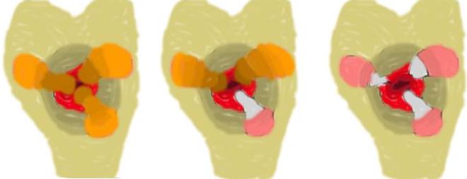
At the end of the procedure the following rhyme should be recalled;
If it looks like a clover
Your trouble is over
If it looks like a Dahlia
It is sure to be a failure
Most surgeons do not insert a tight pack into the anal canal as this causes severe post-operative pain. A soft dressing or a sheet of alginate may be inserted.
 An alginate roll will result in less pain post
haemorrhoidectomy than a parraffin gauze roll. [IWI98]
An alginate roll will result in less pain post
haemorrhoidectomy than a parraffin gauze roll. [IWI98]