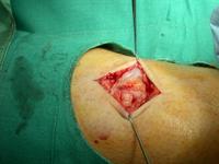
The patient is lying prone.
The posterior aspect of the left calf has been cleaned with an iodine solution
and draped with a green drape. A verticle incision is seen on the posterior
aspect of the calf. This is being held open with skin hooks.
The incision has been carried down through the sub-cutaneous fat and the
deep fascia, which is seen as the white sheet like structure being held by the
skin hook in the upper part of the image. Beneath the deep fascia, I can see
the calf muscles. On the surface of the calf muscles there is a white shiny
structure with tiny blood vessels apparent on its surface. This looks like a
nerve and is most likely the sural nerve.
To the left of the image, I can see the nodular structure that the patient
complained of. This appears to have a similar colour to fat and has a fine
capsule. It appears attached to the nerve in the upper part of the incision.
The incision is on the back or posterior aspect of the calf.
A scar is a healed wound. This is a new incision.
The skin is being held with skin hooks.
The white shiny structure visible in the middle of the wound is the
sural nerve. The Achilles tendon is much bigger than this and
is seen lower down. The tiny blood vessels suppling the nerve can be clearly
seen.
The deep fascia has been divided, it is best seen as the white sheet like
structure being held up by the skin hook in the upper part of the image.
An iodine containing solution (Povidone Iodine) has been used to prepare
the skin. This is evident by the brown colour.
