Image dscn0934 |
| Last updated (17 November 2003). |
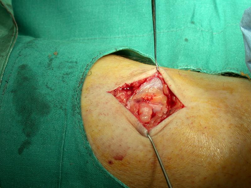
This image shows the back of the left calf. The patient is prone. The left part of the image corresponds to the patients knee. The right part of the image corresponds to the ankle. The upper part of the image corresponds to the patients midline. The lower part of the image corresponds to the lateral aspect of the patients calf.
This image shows the posterior aspect of the patient's left calf. A vertical incision has been made through the skin and the deep fascia. The under surface of the deep fascia is seen as the shiny white sheet like material in the upper part of the wound.
Beneath the deep fascia, there is a white shiny structure with tiny blood vessels on its surface. This structure is superficial to the muscle and has the appearance of a nerve. Given the position of the patient and the incision, this is the sural nerve.
A yellowish nodule is seen attached to the nerve in the upper part of the incision. This nodule appears encapsulated.