The answer
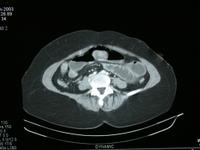
The patient is lying on their back and there are distended loops of
intestine
What the student might say
This is a dynamic CT scan through the mid to lower abdomen. The
patient appears obese. We can see the contrast agent increasing the
density in the blood vessels in the retroperitoneum and in the wall of
the intestine. I cannot read the patients name or the date of birth, but
the scan was performed in June 2003.
The patient is obese, as seen from the thickness of the fat.
The small bowel is distended with fluid and gas, in keeping with
intestinal obstruction.
There is an unusual density sitting in the anterior distended small
bowel loop. This has more the appearance of something lying in the lumen
of the bowel than something growing from the wall of the bowel into the
lumen.
Small print
The patient is supine, this is the usual position for abdominal CT
scans, moreover, the gas/fluid level in the small intestine is
horizontal with the gas uppermost indicating that the patients front is
uppermost.
There is no evidence of an aneurysm on this image.
The appearance in the patient left loin is due to small bowel
distended with fluid. The left kidney is usually superior to this
level.
