Image dscn1199 |
| Last updated (28 October 2003). |

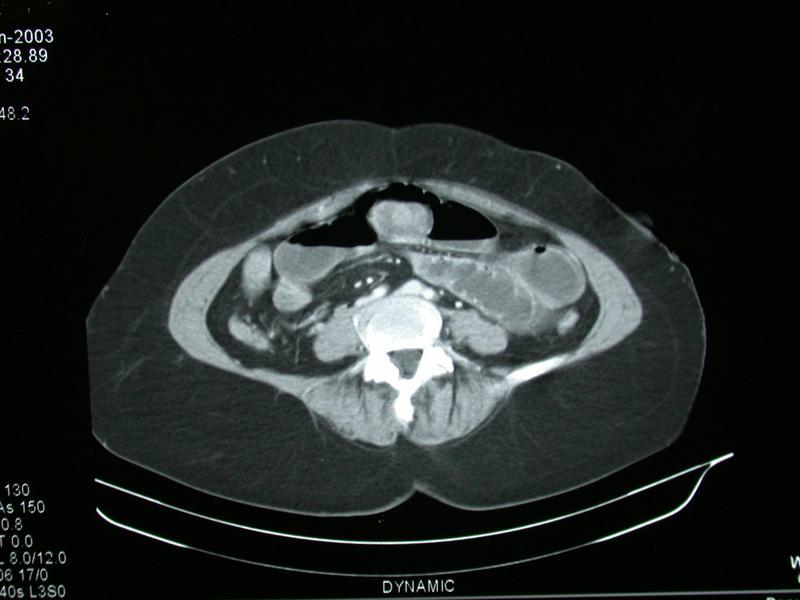
This image is a section taken from the middle to lower abdomen of a CT scan. We know it is the middle to lower abdomen because the upper abdominal organs, the liver and spleen are not visible. It is lower than the middle abdomen because the kidneys are not visible. It is not very low down because the iliac bones are not well seen, only the top of the left iliac crest is visible.
As per usual, the lower part of the image corresponds to the patients back, the upper part the front, the left side of the image corresponds to the patients right hand side and the right side of the image corresponds to the patients left hand side. The image is a slice taken through a patient lying on the scan table with the lower limbs poining out towards you and the head away from you.
The scan is labelled dynamic in the lower part of the image. This indicates that the patient has received contrast, oral and/or intravenous or both. The blood vessels in the retroperitoneum appear bright due to the effects of intravenous contrast, they appear iso-dense with the vertebra. In addition the muscles and wall of intestine appear brighter than usual due to the presence of the contrast agent.
The patient appears overweight, as seen by the large amount of fat.
The small intestine appears to be abnormally distended with fluid and gas. The level of the fluid is running across from left to right with the gas above the fluid, this indicates that the patient is lying supine.
There is an unusual mass lying in the anterior loop of intestine, it appears to be a mass sitting in the lumen of the bowel rather than growing from the wall of the bowel into the lumen.