The answer
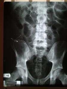
The PFA shows that the previously noted unusual opacity has disappeared.
This confirms the impression that it was the faecolith that caused the
opacity. There is evidence of a midline incision and a surgical drain in the
right iliac fossa.
The most likely course of events at the original operation was that the
patient suffered obstructive appendicitis due to a faecolith becoming
impacted in the appendix. This resulted in a more rapid than usual rate of
progression of the appendicitis and early perforation. The appendix most
likely lay in a retro-caecal position which may have masked some of the
symptoms and signs of appendicitis which would have been more pronounced if the
appendix lay in an intra-peritoneal location. At the time of surgery
the appendix was noted to be perforated and the site of the perforation was
mistaken for the tip of the appendix. The retro-caecal location of the
appendix with severe surrounding inflammation masked the faecolith and the
true tip of the appendix. It is possible that the laparosocpic approach led to
an inferior visualisation of the area where the faecolith and true
appendicular tip lay. After initial recovery, bactria in the faecolith and
residual appendix caused on going inflammation and sepsis, which culminated in
the present admission and surgery.
What the student might say
This PFA thankfully shows that the unusual density is gone, confirming
that it was due to the faecolith. There appears to be a drain in the right
iliac fossa which is secured with a safety pin. Numerous skin clips are seen
in the middle of the abdomen indicating a midline incision. The intestine
appears gassy as it often does in the immediate post operative period due to
an ileus.
The most likely course of events at the original surgery was that the patient
had a perforated appendicitis due to obstruction from the faecolith. At
surgery the perforation in the appendix was mistaken for the appendicular tip
and the true tip and faecolith left in situ. This may have been aggravated by
the retrocaecal position of the appendix and the laparoscopic approach with
poorer vision of the retroperitoneum. Subsequently the faecolith and retained
tip caused further inflammation and sepsis.
Small Print
The unusal opacity is gone
There is a drain in the right iliac fossa
The appearances of the film and the clinical scenario make ileus much more
likely than obstruction. There are no fluid levels seen. The clinical
distinction between ileus and obstruction in a post laparotomy patient is
difficult. If return of bowel function is delayed then obstruction is
considered.
The metal clips seen down the middle of the image indicate a midline
incision. The patient could also have a right iliac fossa incision but this
cannot be seen on the PFA.
Indeed the metallic object in the right iliac fossa is a safety pin. This
is put onto the end of the drain so that the drain will not migrate into the
abdomen. Initially the drain is secured to the skin with a suture, this
prevents the drain from coming out (and moving in). When the drain has done
its job, the suture is remove and the safety pin remains to prevent the drain
migrating into the abdomen. The drain is shortened gradually until it comes
out completely. Thus it can be said that the function of the suture is to stop
the drain coming out and the function of the safety pin is to stop the drain
falling in.
