The answer
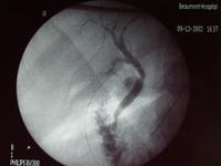
This is a normal per operative cholangiogram.
Note the normal anatomy, the normal calibre of the ducts, the lack of any
filling defects and the normal flow of contrast into the duodenum
What the student might say
This is a per operative cholangiogram. I can see the catheter clipped into
the cystic duct. There is no evidence of biliary dilatation. There are no
filling defects and the contrast is flowing freely into the duodenum. This
looks normal to me.
Small Print
This is not an angiogram. It would be highly unusual practice to perform
an angiogram during an emergency cholecystectomy for a perforated gallbladder.
In any case neither the hepatic arteries nor the branches of the portal vein
look like this when they contain contrast.
This does not show the pelvis of the kidney. It is not a retrograde
pyelogram. The ureter would not normally drain into the intestine and the
branching pattern of the structure seen on the image does not look like a
kidney.
This is not an MRI.
An x-ray of the branches of the portal vein would not be done at the time
of an emergency cholecystectomy for a perforated gallbladder. The branches of
the portal vein do not look like this, the lower parts look wider and the
branching pattern is different. And the portal vein does not drain into the
intestine.
