Image dscn1000 |
| Last updated (28 October 2003). |

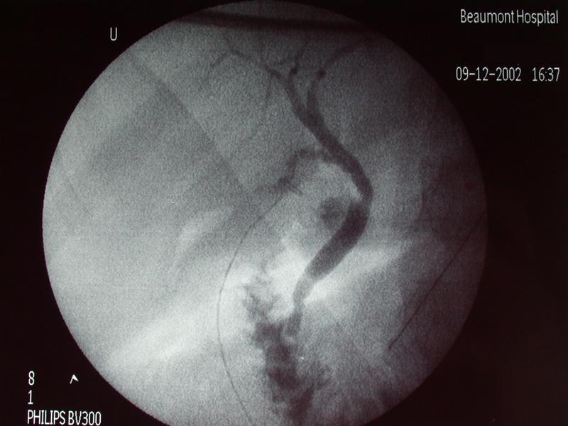
This image shows a per operative cholangiogram. The top of the image points to the patients head. The left side of the image corresponds to the patients right hand side.
This image shows a normal per operative cholangiogram.
The cholangiogram catheter is seen clipped into the cystic duct.
There is normal filling of the intra-hepatic, common-hepatic and common bile ducts. No filling defects are seen. There is flow of contrast into the duodenum. The sphincter of Oddi is in systole during this image and this gives the characteristic inverted meniscus appearance.