| Good Stuff |
| Spot Diagnosis |
| Biliary System |
|
|
|
|
|
|
|
|
|
|
|
|

|
Click on the image to download a larger version |
|
This lady was admitted with a 4 day history of severe upper abdominal pain.
She was tender in the right upper quadrant with a positive Murphy's sign. An
ultrasound of the right upper quadrant confirmed the clinical suspicion of
symptomatic cholelithiasis. There was ultrasonographic evidence of a thickened
gall bladder wall with a single large gallstone seen impacted in Hartman's
pouch.
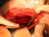
She was initially treated conservatively, but then had to undergo an emergency laparotomy. The image shows the intraoperative findings, what is seen? What complication has occurred? |
Image taken at laparoscopic surgery
An upper midline incision A mini laparotomy Acute gangrenous cholecystitis A hole in the gallbladder |