Image dscn0837 |
| Last updated (28 October 2003). |

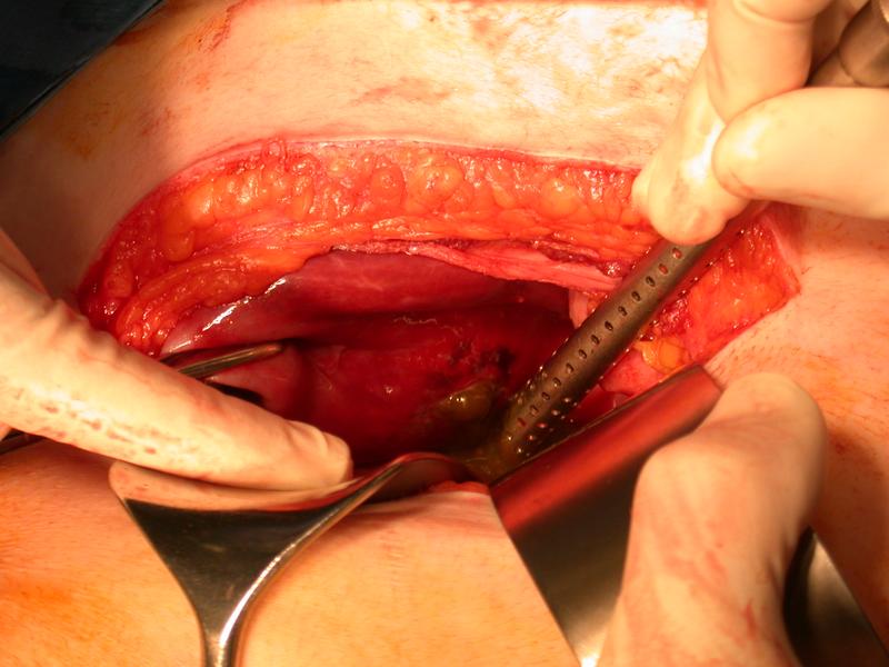
The image is an intra-operative image of the patient's right upper quadrant. The top of the image corresponds to the patients right costal margin. The right part of the image corresponds to the midline of the patients epigastrium.
This image shows an intra-operative view of a patient who has had a right subcostal (Kocher) incision. This type of incision is most commonly done for open cholecystectomy.
There are two retractors in the lower part of the incision. A sponge holding forceps to the left of the image appears to be holding the fundus of the gallbladder.
The anterior and posterior layers of the rectus sheath are seen anterior and posterior to the rectus abdominus muscle in the upper part of the incision. The liver is seen in the upper part of the abdomen beneath the fascia, with the gallbladder attached to its under surface. The tip of the sucker is adjacent to a large hole in the gallbladder. Bile coloured material is issuing from the hole. Surrounding the hole the wall of the gallbladder appears black, suggesting infarction.