The answer
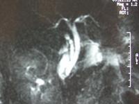
This image is a magnetic resonance cholagio-pancreatogram (MRCP). It shows no
evidence of a distal obstruction or a defect in the common bile duct. There is
a long cystic duct stump and there appears to be a puddle of fluid in contact
with the end of the cystic duct stump.
What the student might say
This image is a magnetic resonance cholangio-pancreatogram (MRCP).
It shows a long cystic duct remnant with puddle of fluid associated with
the end of the stump, this is probably the site of the leak and corresponds
nicely with the laparoscopic image.
Small Print
HIDA scans are nuclear medical scans which examine the biliary system.
They usually appear spotty due to the effects of the gamma rays from the
administered radio pharmaceutical on the gamma camera. The spots where there is
radioactivity are usually seen as black.
This is not a 3d reconstruction from ultrasound or CT. 3d reconstruction
from CTs is done particularly in vascular imaging. It would be difficult to get
enough contrast in the bile duct to get a nice 3d reconstruction from a CT.
If you were going to directly inject contrast into the biliary system then you
can usually get nice images with fluroscopy.
No this is not an EM picture of thread worms.
