Image dscn1104 |
| Last updated (28 October 2003). |

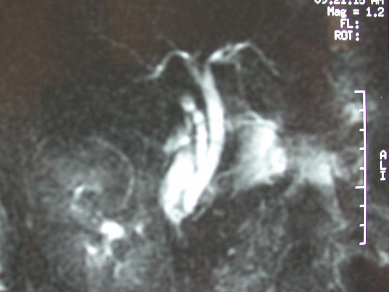
This image shows an MRI examination of the bilary system and pancreas, a so called magnetic resonance cholangio-pancreatogram (MRCP).
The biliary system appears full, as it may do in cases of distal obstruction, or following cholecystectomy. No gallbladder is visible probably indicating cholecystectomy. There is no evidence of a filling defect in the distal bile duct. Extending from the lower part of the bile duct towards the patients left hand side, a small main pancreatic duct is visible.
A long cystic duct remnant is visible, there appears to be some fluid beside the end of the cystic duct remnant.