Biliary System

Last updated (28 October 2003)

Leaky Bile Click on the image to download a larger version
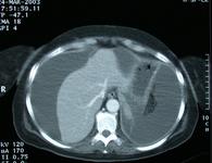
This image is from an emergency scan taken in a lady 3 days post laparoscopic cholecystectomy.
What sort of scan is it?
What does it show?
A MRI
A dynamic CT scan
Computerised reconstruction of an Ultrasound scan
Normal abdominal CT scan
Improve your X-Ray reading skills

The answer

This ia a dynamic CT scan taken through the upper abomen. It shows considerable ascitis which is compressing the liver and the inferior vena cava.

What the student may say

This image shows an axial section of a CT scan taken through the upper abdomen. The patient has been given intra-venous contrast.

There is evidence of a large amount of intra-abdominal fluid. It would appear that the fluid is compressing the liver and inferior vena cava.


Adrian P. Ireland