Image dscn1087 |
| Last updated (28 October 2003). |

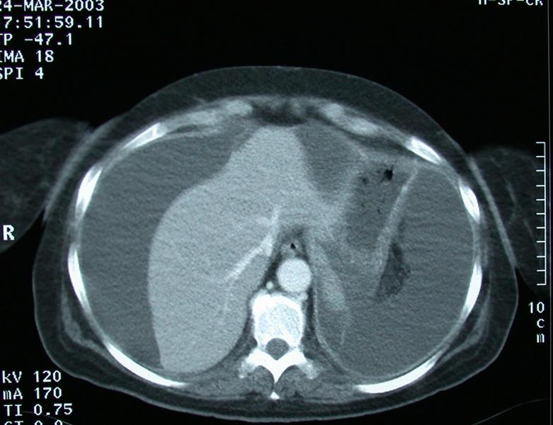
This section is taken from a CT scan through the upper abdomen. IV contrast has been administered, as seen by the bright signals over the aorta and portal vein, the liver is also brighter than normal (enhanced). There is no evidence of contrast in the stomach so oral contrast has probably not been given.
There is a considerable amount of ascitis present.
The Inferior vena cava is not visible. The cava may not be visible because it is absent, or it has fibrosed due to previous inflammation. In this pariticular patient the association of considerable ascitis with difficulty in seeing the cava makes twisting of the liver due to the ascitis and kinking of the cava the most likely caused, this may result in haemodynamic alterations with hypotension etc. Rapid accumulation of the acitis is necessary to cause this.
Kinking of the IVC due to twisting of the liver from rapidily accumulating ascitis due to a bile leak post cholecystectomy is termed the Waltman-Walters syndrome.