Image composite |
| Last updated (28 October 2003). |

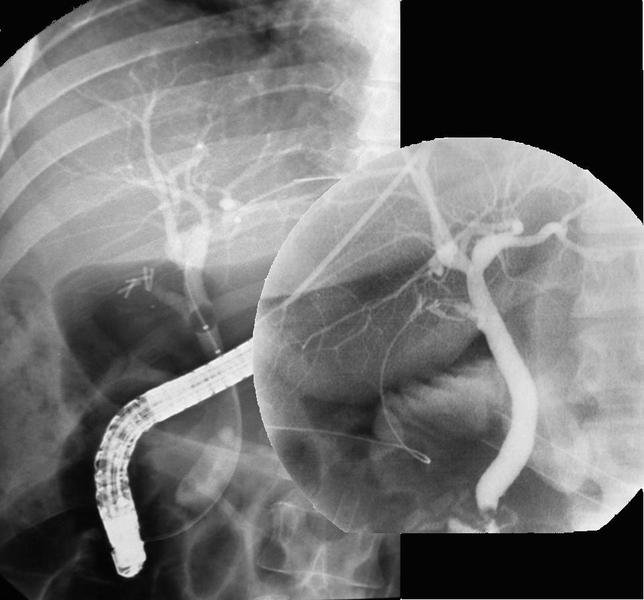
This is composite of two images.
The image on the left is an X-ray of the right upper quadrant of the patients abdomen. The top of the the left image corrsponds to the bottom of the patients right lung. The right part of the left image corresponds to the lateral part of the right upper quadrant of the abdomen. The left part of the left image corresponds to the midline (see the vertebra). The lower part of the left image corresponds to the disc between L2 and L3.
The circular image on the right is a peroperative cholangiogram. Further information about this image is given here.
This image is taken at the time of an endoscopic retrograde cholangio-pancreatogram (ERCP). The large radio-opaque tube like structure is the endoscope as it passes through the esophagus and stomach into the duodenum.
Numerous clips are seen in the region of the porta hepatis, suggesting previous surgery in this location.
Contrast has been injected into the bile duct and the dark area in the lower duct corresponds to lack of filling with contrast due to the presence of a stone.
The dark area higher up the image corresponds to a balloon which has been inflated in the duct. The balloon is being used to pull the stone back out of the duct into the duodenum.
The inset on the right is the peroperative cholangiogram taken at the time of cholecystectomy. Compare the appearaces then with that seen on the ERCP.