Image dscn0981 |
| Last updated (28 October 2003). |

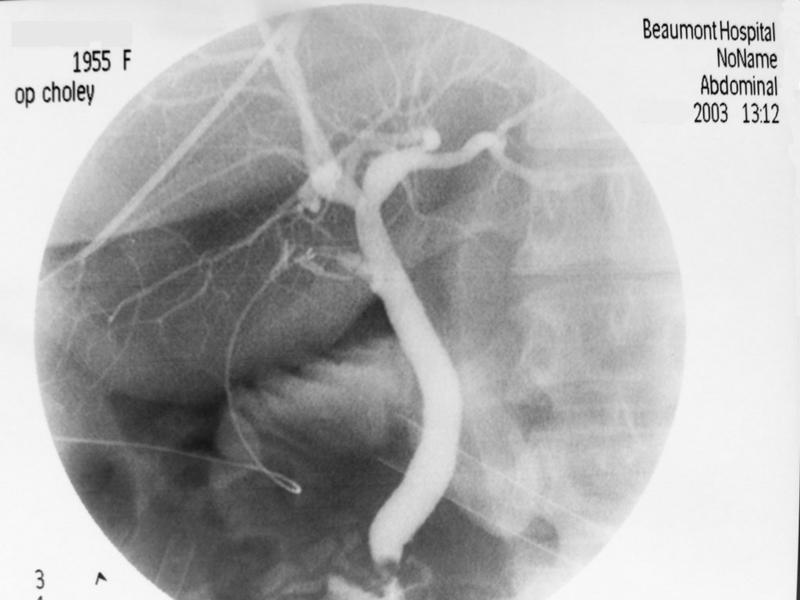
This is a per operative cholangiogram. The top of the image is at T10. The bottom of the image is at L3. The right part of the image corresponds to the patients midline. The left part of the image corresponds to the patients right hand side.
This is a peroperative cholangiogram. The cholangiogram catheter is seen clipped into the cystic duct. A laparoscopic port is visible in the right lower part of the image. A further smaller laparoscopic port is visible at the 7 o clock position. This cholangiogram was taken at a laparoscopic cholecystectomy.
A filling defect is seen in the lower part of the bile duct. The caliber of the bile duct appears normal. There is normal flow of contrast into the duodenum.