The answer
There are many possibilities, ranging from open exploration and placement
of a T tube to post operative MRCP with ERCP if that is abnormal.
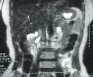
The image shows a coronal section from a magnetic resonance cholangio
pancreatogram. The filling defect in the lower end of the bile duct
persists.
What the student may say
This is a controversial area and there is no simple answer. I would ask
the opinion of the consultant surgeon caring for the patient.
The decision would depend on the size of the stone and the duct and local
expertise. Unsuspected stones are usually small in a small duct, particularly
if the ultrasound has been done close to the time of surgery as ultrasound
will identify large ducts easily. If further intervention is planned it would
be prudent to ensure that the filling defect seen on the cholangiogram is a
stone and not for instance a gas bubble, i.e. a false positive.
If the stone is large and the duct is large then it may be best to proceed
to exploration of the duct. Exploration is most often done as an open
operation but it may be accomplished laparoscopically if the surgeon is expert
in this area. However, unless the surgeon is doing alot of it, he/she may not
be able to maintain sufficient expertise.
At open surgery the duct may be explored from above (through an opening in
the duct made in the edge of the lesser omentum), or from below (through the
ampulla via a duodenotomy). Most surgeons would usually do the exploration
from above. The risk with the from below
approach is damage to the pancreatic duct with postoperative pancreatitis. A
formal surgical sphincteroplasty is usually performed so there is a low risk
of subsequent stenosis. Widening the opening from the duodenum into the bile
duct will aid in the the passage of any residual solid material into the
duodenum. A very wide opening may increase the chances of cholangitis though.
If the duct is opened from above then following removal of
the stone it is best to do a completion choledochoscopy to ensure that there
are no further stones visible. A latex T tube is placed in
the common duct and brought out through the skin. The patient can have a
tubogram about 10 days post operatively to check that there are no residual
stones prior to pulling the tube out.
If the duct and stone are small, then about 50% will pass without
intervention. Thus it may be reasonable to not explore the duct in
anticipation that the patient will pass the stone spontaneously.
If the duct is not explored and a suspected stone has been identified on
the cholangiogram, then post operative follow up is vital. The patient should
be monitored for pain and abnormal liver function tests. The duct should be
evaluated to ensure that the stone has passed. This evaluation can be in the
form of a magnetic resonance cholangio pancreato-gram (MRCP), an ERCP or a
tubogram via a catheter placed in the common duct via the cystic duct at the
time of surgery.
If the duct evaluation shows that the stone has not passed then it should
be removed. The advantage of ERCP in this situation is that open surgery can
be obviated. The disadvantages are
- It may not be possible on ERCP to find the duct.
- It may not be possible on ERCP to remove the stone.
- The patient may suffer a serious complication in the form of bleeding,
perforation or pancreatitis.
- The patient may develop papillary stenosis following papillotomy and
thus develop further problems in the future e.g.. recurrent common bile duct
stones and cholangitis.
If the ERCP fails then the patient may have to undergo further surgery.
The image shows an MRCP with persistence of the filling defect seen on the
per operative cholangiogram. This indicates that the stone was not extracted at
the time of surgery and the scan was performed to see if the stone had passed,
it had not, so the patient most likely was referred on for ERCP.
Small print
As usual, when there are lots of options, there is lots of controversy. If
one particular option was clearly superior, the other options would no longer
be options and there would be no controversy.
Careful clinical follow up is not a good option, if you have identified
the stone you will have to do some further imaging to see what has happened to
it. It is standard practice in many centres not to do a cholangiogram at the
time of a cholecystectomy and in essence those patients undergoing
laparoscopic cholecystectomy without per operative cholangiography who have
unsuspected common bile duct stones are followed clinically but this is not
equivalent to the situation where you have done a cholangiogram and discovered
an unsuspected stone and you follow the patient clinically.
Laparoscopic extraction is possible, but gaining the skill to do it is
difficult. It would seem that the surgeon doing it would have to be doing it
quite regularly to get really good at it.
Open surgery is an option, but this is not attractive if the size of the
duct is small. There is also a definite false positive rate on
cholangiography. As for any operation, the surgeon must discuss the
possibility of an open operation with the patient beforehand. If the patient
undergoes supra-duodenal antegrade exploration then a T-tube is always placed
in the duct through the opening. Bile is very slippy and even if the hole in
the duct is approximated with numerous fine sutures, bile will continue to
leak out even through the holes made by the sutures. The T-tube helps to drain
the bile, it also stents the duct to reduce the chance of it narrowing
afterwards and it offers an opportunity to check for residual stones via a
tubogram. If there are residual stones then the T-tube is left in situ until
a mature track is formed. This track can then be used to extract the
stones.
Post operative ERCP is an option. But it may not be necessary, if the
cholangiogram was a false positive or the stone passes spontaneously. In
addition there is a low but definite morbidity and mortality associated with
ERCP.
Some surgeons advocate placement of a fine bore tube in the bile duct if
the per operative cholangiogram is positive and the duct is small. This
facilitates repeated imaging to rule out a false positive result. It will also
offer objective evidence that the stone has passed without having to do an
ERCP. If the stone does not pass the tube will act as a vent in the bile duct to
reduce the chances of cholangitis.
A further alternative is to proceed with the laparoscopic operation and do
an MRCP in the post operative period to check the duct. If the duct is clear
the patient may be followed clinically and with liver blood tests. If the duct
is not clear on MRCP then they may proceed to ERCP.
Some surgeons do not perform a per operative cholangiogram at the time of
cholecystectomy. This saves time and money. They will not have to worry about
discovering an unsuspected common bile duct stone. The patient will either
pass the stone spontaneously or return with symptoms due to the unsuspected
stone. If they return with symptoms then further evaluation and treatment may
be necessary.
