The answer
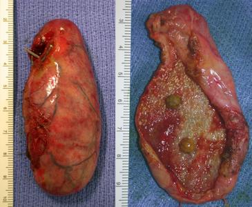
The gallbladder has been removed intact, the image on the right shows the
gallbladder opened. There are in fact gallstones present, two are seen sitting
on the gallbladder mucosa. The gallbladder wall appears markedly thickened in
keeping with cholecystitis. There are multiple white plaques present which
represent cholesterolosis of the gallbladder wall.
What the student may say
I can see an intact gallbladder on the left, the superficial blood vessels
appear injected suggesting inflammation.
To the right the gallbladder has been opened, two 4 mm gallstones are
sitting in the gallbladder. The gallbladder wall is thickened and numerous
white plaques which are typical of cholesterolosis are visible.
The white appearance is due to the accumulation of large amounts of
cholesterol in foamy macrophages.
Contributing factors to missing the gallstones on ultrasound may have been
the fact that they are small and few in number.
Small print
Cholesterolosis is a curiosity that may be seen upon opening the
gallbladder in patients with cholecystitis. It does not have any particular
significance. Most surgeons would routinely send an excised gallbladder for
histological examination. Recently, some have questioned this as a waste of
money and resources and suggest that only suspicious gallbladders should be
submitted. Histological examination would be particularly important if there was
any evidence of macroscopic abnormalities.
There is no evidence of perforation of the gallbladder on the left. It
appears inflated with bile, etc. If a perforation does occur, gallstones may
spill into the peritoneal cavity and if they are not retrieved they may
subsequently cause trouble.
The operative finding agree nicely with the radiological diagnosis of
cholecystitis, the gallbladder wall is thickened and there is cholesterolosis.
But, the operative findings show gallstones which were missed on the
ultrasound. Therefore the patient did not suffer acalculous
cholecystitis. Ultrasound is a good test, however, no test is 100% accurate,
there will be the occasion when it is wrong. In this particular patient, the
fact the gallstones are small and few in number possibly contributed to the
inaccuracy of ultrasound.
The gallbladder wall appears markedly thickened on this image.
