Image composite2 |
| Last updated (28 October 2003). |

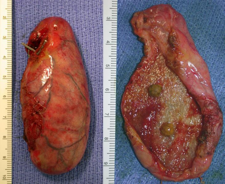
This image is a composite of two views of a gallbladder that has been excised from the patient. The fundus of the gallbladder is seen to the bottom of the image and the cystic duct end to the top.
In the left hand image the gallbladder is shown intact, clips are seen on the cystic duct and artery. The peritoneal covered surface of the gallbladder is to the right and the part of the gallbladder that was in contact with the liver is shown to the left.
In the image to the right the gallbladder has been opened through the part that was in contact with the liver. The clips on the cystic duct and artery have been removed. Two gallstones with a diameter of 4 mm are seen sitting on the wall of the gallbladder. The mucosa of the gallbladder appears to have white patches on it. These are macroscopic evidence of cholesterolosis which is due to the accumulation of a large amount of cholesterol in foamy macrophages.
The gallbladder wall appears markedly thickened on the image to the right.