The key to understanding the common colonic resections for cancer is the anatomy of the blood supply to the colon. Resection of the tumor is accomplised en-bloc with resection of the draining lymph nodes. These lymph nodes are arranged along the blood vessels supplying the colon. When the lymph nodes and blood vessels are resected, all of the colon supplied by the blood vessels needs to be resected.
The blood supply to the colon comes from the superior and inferior mesenteric arteries. Part of the rectum is supplied by the internal iliac system and this anastamoses with the lower branches of the inferior mesenteric system. At the top end the branches of the superior mesenteric artery to the proximal bowel anastamose with branches from the coeliac axis.
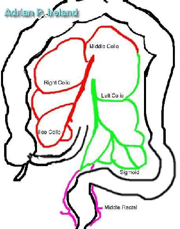
The superior mesenteric artery gives the middle, right and ileo-colic arteries. There is a marginal artery close to the mesentric border of the colon which connects these and via a communication of variable completeness to branches from the inferior mesenteric system in the region of the distal third of the transverese colon to the splenic flexure.
The ileo colic artery is the main termination of the superior mesenteric artery. It supplies the terminal ileum, the appendix, caecum and proximal ascending colon. Its marginal branches join those of the right colic artery.
The right colic artery supplies the ascending colon. Its ascending marginal branches supply the hepatic flexure and proximal transverse colon. The distal marginal branhes join with the marginal branches of the right branch of the middle colic artery.
The middle colic artery arises close to the origon of the superior mesenteric artery in the root of the transverse meso-colon. It quickly bifurcates into right and left branches the right branch joins with the marginal branches of the right colic artery in the region of the hepatic flexure and the left with the marginal branches of the left colic artery in the region of the splenic flexure.
The inferior mesentric artery supplies the descending colon, sigmoid colon and recto-sigmoid junction. The first main branch is the left colic artery which supplies the descending colon by bifurcating into upper and lower branches. The upper branch ascends as a marginal artery to anastamose with the terminal branches of the left branch of the middle colic artery. The lower branch descends as a marginal artery to anastamose with marginal branches of the sigmoid branches of the inferior mesenteric artery.
After the inferior mesenteric artery has given off the left colic artery it continues down giving off numerous branches to the sigmoid colon. These form archades and ansatamoses with each other. The most distal branches continue down to supply the upper rectum.