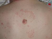
The clinical suspicion can be readily confirmed by a punch biopsy.
The first thing to do is a full history and clinical examination. In
particular I would enquire about a past history of any cancers, the
family history, occupation (indoors or outdoors) and history of sunburn.
On examination, I would carefully look for evidence of metastatic
disease in particular in the adjacent lymph node basins, the
supraclavicular fossa, both axillae and in the mid lateral trunk. I
would carefully inspect the skin between the lesion and these lymph node
basins to look for in-transit metastases.
There is no blood test that would be of any particular interest.
Detailed scanning should be avoided untill the suspected diagnosis is
confirmed. A CXR may be worth while however.
The diagnosis is most readily confirmed by a punch biopsy. An excisional
biopsy could be considered if the lesion was smaller but in this case
excision would not be feasible without embarking on elaborate
reconstruction.
Skin scrapings (and nail clippings) are usually performed to check for fungal
infection.
An MRI may be used to stage melanomas and other tumors but is usually not used
to confirm the diagnosis. The most important modality to confirm the nature of
an abnormality is tissue diagnosis which depends on tissue.
Excisional biopsy could be considered but the large size of the lesion means
that primary closure would not be possible and the defect would then require
closure with some more elaborate means. Following definitive diagnosis with
tissue then a larger excision and further elaborate means again would be needed
to cover the defect.
