Image dscn0650 |
| Last updated (19 November 2003). |

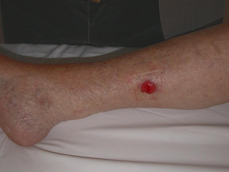
This image shows the postero-medial aspect of the patients right calf. The right part of the image corresponds to the patients knee. The left part of the image corresponds to the patients ankle. The patients heel is seen in the lower left part of the image.
The patient is lying supine with the right lower limb externally rotated.
The major abnormality is a raised cherry red lesion over the lower margin of the medial head of the gastrocnemius muscle.
Small superficial veins are visible and there is some brown discolouration noted over the anterior ankle area suggesting hemosiderin deposition. A venous flare or telangictasia is visible just posterior to the medial malleolus These features raise the possibility of venous insufficiency. However, the condition of the skin in the calf on this aspect of the leg is healthy, apart from the raised cherry red lesion.
An imprint of a dressing is visible as a rectangular indentation around the lesion. Of note, the skin where the lateral aspect of the dressing indentation is shows evidence of bruising, possibly due to trauma from the sticky tape used to hold the dressing in place.
The lesion is approximately 2.5 cm in diameter. It is elevated above the surface of the skin. The lesion appears to be well vascularised.