Image shastri02 |
| Last updated (28 October 2003). |

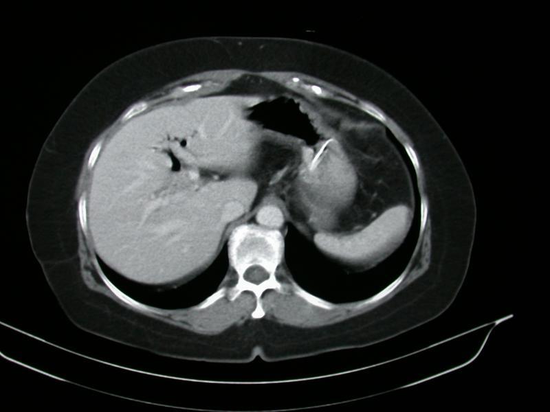
This is a section taken from a CT scan through the upper abdomen The top of the image corresponds to the patients front. The left of the image corresponds to the patients left hand side. The image is a slice taken through a patient lying on the scan table with the lower limbs poining out towards you and the head away from you.
The patient appears overweight, note the substantial fat.
This is a contrast enhanced or dynamic CT scan as seen by the increased density over the aorta and vena cava, which lie anterior to the vertebral column. In addition the spleen and liver are brighter than normal.
There is a bright linear abnormality in the stomach, which would correspond to a naso-gastric tube.
There is an abnormal gas pattern visible in the liver. This gas pattern follows a branching patern and could be in either the portal vein or the biliary tree, it is most likely in the biliary tree (gas in the portal vein may occur rarely in critically ill patients with gas forming organisms in the portal circulation).