Image dscn0906 |
| Last updated (28 October 2003). |

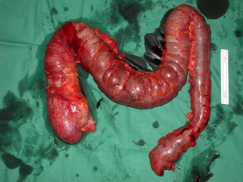
This image shows the entire colon which has been removed from the patient. The colon appears to be orientanted anatomically with the caecum to the lower left corner of the image and the hepatic flexure in the left upper left corner. The splenic flexure is in the right upper corner of the image and the recto-sigmoid junction in the lower middle part of the image.
This image shows a colon that has been removed from the patient.
Very little mesentry is attached to the colon, suggesting that no attempt to remove draining lymph nodes was made.
Only a tiny segment of ileum is included, suggesting that no attempt to remove draining lymph nodes in the ileo-colic region was made. Such an attempt would have mandated excision of part of the ileo-colic artery and further resection of ileum.
The right and transverse colons are markedly distended. The distal descending colon and sigmoid appear of normal calibre, however, no obvious obstructing lesion is visible.
The surface of the distended colon shows inflammation with adherent fibrinous material. Many of the blood vessels appear distended. There is a patch of dusky material best seen in the mid transverse colon, suggesting underlying mucosal infarction.
The appearances are those of a toxic colitis.