Image dscn0957 |
| Last updated (28 October 2003). |

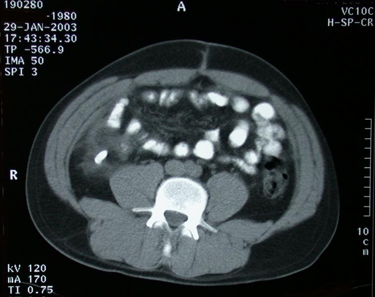
The is a section taken from an CT scan through the mid to lower abdomen.
See notes on orientation of CT scans.
This section shows the mid to lower abdomen. We know this because we are below the kidneys and above the iliac crests.
There is a track leading from skin in the anterior abdomen in the midline into the abdominal wall. This is in keeping with a previous wound. This does not appear to be the normal scar seen in the vicinity of the umbilicus. But would be in keeping with previous insertion of a laparoscopic port.
The patient has taken oral contrast. We know this because the intestine is isodense with the vertebra indicating the presence of contrast. However, there is no evidence of contrast in the major blood vessels in the retroperitoneum.
To the patients right side there is an unusual density which lies behind the ceacum or ascending colon. Surrounding this density there is evidence of stranding of the fat, indicitive of inflammation.