Image dscn0840 |
| Last updated (28 October 2003). |

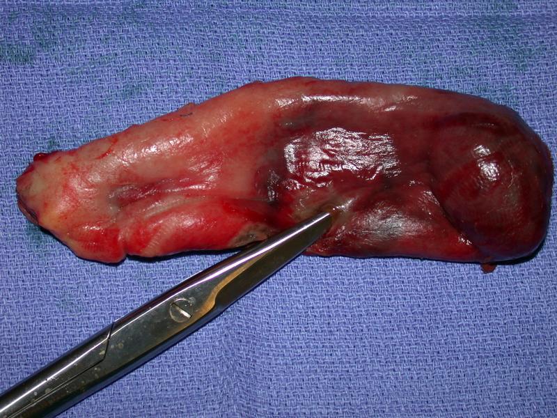
The image shows a gallbladder which has been excised from the patient.
The fundus is to the right of the image and Hartman's pouch to the left.
The inferior surface of the gall bladder is shown, (covered with peritoneum).
This images shows an operative specimen of a gall bladder.
There is a large mass in the fundus of the gallbladder which probably represents a single large gallstone.
About half way down the gallbladder there is a hole which is indicated by the tip of the scissors, this corresponds to the site of perforation.
The gallbladder around the hole shows signs of necrosis, it is green and dull in comparison to the adjacent tissue.