The answer
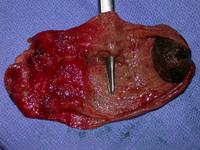
The inside of the gallbladder is show. A single large gallstone is lying
in the fundus. There is severe inflammation in the area of
Hartman's pouch. Just above Hartman's pouch
there is a hole in the gallbladder indicated by the tip of the scissors.
The student may say
I can see the inside of the gallbladder. To the right a single large
gallstone is seen lying in the fundus. To the left there is evidence of severe
inflammation in Hartman's pouch.
It is most likely that the stone seen on the right was impacted in
Hartman's pouch, (as seen on ultrasound). The inflammation would support that.
The perforation seen just above Hartman's pouch would correspond to a site in
the gallbladder wall that was stretched and thereby liable to pressure
necrosis.
Small print
The gall stone is seen in the fundus of the gallbladder.
There is no macroscopic evidence of cholesterolosis here. Cholesterolosis
appears as multiple white spots on opening the gallbladder.
Rokitansky Aschoff sinuses are only seen on microscopy.
