Image dscn1092 |
| Last updated (28 October 2003). |

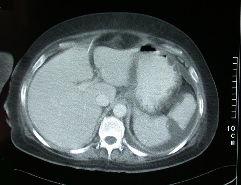
This image shows a section from an abdominal CT taken through the upper abdomen. The top of the image corresponds to the front of the patient. The left side of the image corresponds to the patients right hand side.
This image shows a section from an abdominal CT scan taken through the upper abdomen. We know it is the upper abdomen, because the liver and spleen are visible.
Both intravenous and oral contrast have been given. The effects of the intra-venous contrast are seen by the high density seen in the aorta and inferior vena cava, the increased density seen in th elver and spleen. Oral contrast can be seen in the stomach.
A linear density seen just to the right of the midline is in keeping with the presence of a drain.
A segment of the spleen is not enhancing suggesting the presence of a wedge shaped splenic infarct.
A small amount of free fluid is visible, especially behind the spleen.